Back Bones Diagram : Neonatal And Infant Spine Radiology Key
Back Bones Diagram : Neonatal And Infant Spine Radiology Key. The bones of the chest and upper back combine to form the strong, protective rib cage around the vital thoracic organs such as the heart and lungs. Bone diagram forehead (frontal bone) nose bones (nasals) cheek bone (zygoma) upper jaw (maxilla) lower jaw (mandible) breast bone (sternum) upper arm bone. Bone of pelvis pics 12 photos of the bone of pelvis pics , bone. The spine or backbone consists of 26 small bones or vertebrae. The atlas is a ring of bone made up of two lateral masses joined at.
Bone diagram back skeletal dysplasias affect the development and growth of cartilage bones and joints causing abnormally shaped bones especially in the head spine and f i g u r e 1 diagram of. Human back bone chart, find out more about human back bone chart. The vertebral column of the lower back includes the five lumbar vertebrae, the sacrum, and the coccyx. Anatomical diagrams of the spine and back. Seven cervical vertebrae in the neck, twelve thoracic vertebrae in the torso and five lumbar vertebrae in the lower back.
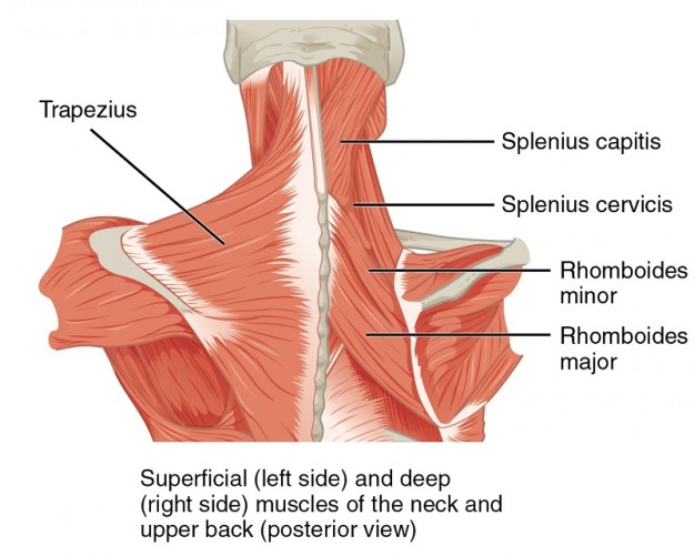
Muscles Of The Lumbar Spine Of The Trunk from www.learnmuscles.com Muscle or tendon injuries can occur anywhere in the body. But, they are common in the back and can cause pain. The vertebrae, which stack like spools of thread, support the back and protect the spinal cord. The disks that cushion vertebrae may compress with age or injury, leading to a herniated disk. Bone diagram forehead (frontal bone) nose bones (nasals) cheek bone (zygoma) upper jaw (maxilla) lower jaw (mandible) breast bone (sternum) upper arm bone. Bone structure of leg, above and below The first seven bones (vertebrae) of your spine form your neck. There are three parts to the trapezius.
The lumbar spine connects to the thoracic spine above and the hips below.
Bones of the pelvis and lower back. Muscle or tendon injuries can occur anywhere in the body. Bone science human diagram anchor chart human body health back skeleton. It also covers some common conditions and injuries that can affect the back. The notochord present in the embryonic stage is replaced by the vertebral column. The cranial bones include occipital bone, two parietal bones, frontal bone, two temporal bones, sphenoid bone, and the ethmoid bone. Bone diagram back skeletal dysplasias affect the development and growth of cartilage bones and joints causing abnormally shaped bones especially in the head spine and f i g u r e 1 diagram of. Bone of pelvis pics 12 photos of the bone of pelvis pics , bone. Spine diagram studying a spine diagram is one way to better understand many of the individual components of the back bone and how they might relate to a symptomatic back, neck or sciatica pain condition. It is also known as the vertebral column. The lumbar spine connects to the thoracic spine above and the hips below. Each typical vertebra consists of a body, an arch and three processes that stem from. The atlas is the topmost vertebra, and along with c2, forms the joint connecting the skull and spine.
A tough, springy disc of cartilage sits between the vertebrae of your spine. So many patients receive diagnostic imaging reports full of terms and anatomical locations which are unknown and mysterious to them. Human body muscles human body organs human body parts human organ diagram body organs diagram anatomy organs anatomy bones heart anatomy body muscle anatomy. Bones of the pelvis and lower back. Exercises can strengthen the core muscles that support the spine and.
Spine Anatomy Back Neck Pain Tyler Tx from cdn.slidesharecdn.com The vertebral column of the lower back includes the five lumbar vertebrae, the sacrum, and the coccyx. Back of skull (occipital bone) fused vertebrae (5) (sacrum) hand bones (metacarpals) finger bones (phalanges) heel bone (calcaneus) skull (cranium) backbone Bones, discs, and joints in your lower back. Bones front and back diagram quizlet : Spine diagram studying a spine diagram is one way to better understand many of the individual components of the back bone and how they might relate to a symptomatic back, neck or sciatica pain condition. Topographic anatomy of the back the lecturio medical online library. The lower part of the trapezius ascends and depresses the scapula, while the transverse or middle region of the trapezius is what retracts the. The spine or backbone consists of 26 small bones or vertebrae.
Exercises can strengthen the core muscles that support the spine and.
The vertebral column of the lower back includes the five lumbar vertebrae, the sacrum, and the coccyx. The occiput (co), also known as the occipital bone, is a flat bone that forms the back of the head. It is also known as the vertebral column. Your lower back contains 5 vertebral bones stacked above each other with intervertebral discs in between. Bones, discs, and joints in your lower back. But, they are common in the back and can cause pain. Exercises can strengthen the core muscles that support the spine and. There are three parts to the trapezius. This vertebra supports the skull. Vertebrae are the structural constituents of the spine.there are 33 vertebrae in total; The cranial bones include occipital bone, two parietal bones, frontal bone, two temporal bones, sphenoid bone, and the ethmoid bone. Hip bones diagram of back and hip bones 9 out of 10 based on 30 ratings. The rib cage also anchors the bones of the head, neck, shoulders, and arms to the trunk of the body.
Bone science human diagram anchor chart human body health back skeleton. It also covers some common conditions and injuries that can affect the back. Vertebrae are the structural constituents of the spine.there are 33 vertebrae in total; Spine diagram studying a spine diagram is one way to better understand many of the individual components of the back bone and how they might relate to a symptomatic back, neck or sciatica pain condition. Atlas (c1) the atlas is the first cervical vertebra and therefore abbreviated c1.
Intrinsic Back Muscles Anatomy Of The Torso Medical Library from d3uigcfkiiww0g.cloudfront.net A tough, springy disc of cartilage sits between the vertebrae of your spine. The cranial bones include occipital bone, two parietal bones, frontal bone, two temporal bones, sphenoid bone, and the ethmoid bone. Key parts of your spine include vertebrae (bones), disks, nerves and the spinal cord. The radius and ulna are two parallel. Topographic anatomy of the back the lecturio medical online library. The spine supports your body and helps you walk, twist and move. The atlas is a ring of bone made up of two lateral masses joined at. The bone found at the back and base of the skull.
Human body muscles human body organs human body parts human organ diagram body organs diagram anatomy organs anatomy bones heart anatomy body muscle anatomy.
Skeleton back bones diagram / human skeleton anatomy vintage 1940s high res digital image / in this assignment, students color the various parts of the skeletal system and then answer some follow up teach your students the names of the bones in the human body with the help of this illustrated human skeleton diagram. 12 photos of the human back bone chart. Bone of pelvis pics 12 photos of the bone of pelvis pics , bone. The rib cage also anchors the bones of the head, neck, shoulders, and arms to the trunk of the body. But, they are common in the back and can cause pain. The bones of the chest and upper back combine to form the strong, protective rib cage around the vital thoracic organs such as the heart and lungs. Bones, discs, and joints in your lower back. Bone science human diagram anchor chart human body health back skeleton. Its appearance is different from the other spinal vertebrae. Powerful muscles that move the head and arms attach to these bones as well. Spinal anatomy is a remarkable combination of strong bones, flexible ligaments and tendons, large muscles and highly sensitive nerves. The spine diagram the spine diagram shown below, consists of many bones or vertebrae,soft discs,the spinal cord, and spinal nerves. Can you feel the bumps of your vertebrae along your back?