Abdominal Anatomy : 399 Abdominal Anatomy Muscles Photos Free Royalty Free Stock Photos From Dreamstime - • in this module, we will explore basic abdominal anatomy identifiable with common imaging modalities.
Abdominal Anatomy : 399 Abdominal Anatomy Muscles Photos Free Royalty Free Stock Photos From Dreamstime - • in this module, we will explore basic abdominal anatomy identifiable with common imaging modalities.. The abdomen contains all of the digestive. This mri abdomen axial cross sectional anatomy tool is absolutely free to use. We will wrap up with an overview of several abdominal diseases that might all present themselves. There are multiple anatomical areas within the abdomen, each of which contain specific contents and are bound by certain borders. The abdominal region is supported by the anterior and posterior abdominal wall that supports the viscera and maintains the posture where there's no bony support.
The anterolateral abdominal wall formed of 4 layer skin, fascia, muscles, and peritoneum. The abdomen contains all of the digestive. • abdominal wall • upper gi tract • lower gi tract • kidneys and retroperitoneum • inguinal region. This muscle forms the anterior and lateral abdominal wall. A good amount of area is covered by the abdominal wall.
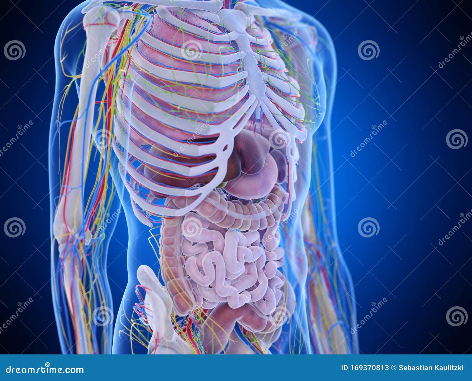
The Abdominal Anatomy Stock Illustration Illustration Of Biology 169370813 from thumbs.dreamstime.com Understanding abdominal anatomy and physiology is essential to understanding the human body as a whole. The anterolateral abdominal wall formed of 4 layer skin, fascia, muscles, and peritoneum. The abdomen (colloquially called the belly, tummy, midriff or stomach) is the part of the body between the thorax (chest) and pelvis, in humans and in other vertebrates. Divided into 9 regions by two vertical and two horizontal imaginary planes. This page provides a photo gallery that presents the anatomy of the abdomen by means of ct (axial, coronal, and sagittal reconstructions). There are some harder ones such as hanging leg raising and body weight. • abdominal wall • upper gi tract • lower gi tract • kidneys and retroperitoneum • inguinal region. Chapter 3 anatomy the anatomy of the et system is related to function and developmental anatomy and is associated with the high rate of otitis fastest abdominal insight engine.
The abdomen (colloquially called the belly, tummy, midriff or stomach) is the part of the body between the thorax (chest) and pelvis, in humans and in other vertebrates.
• in this module, we will explore basic abdominal anatomy identifiable with common imaging modalities. A collection of articles covering abdominal anatomy, including abdominal wall anatomy and abdominal cavity anatomy. The abdomen contains all of the digestive. We'll identify as many organs as we can. The anterolateral abdominal wall formed of 4 layer skin, fascia, muscles, and peritoneum. Sciency root words make anatomical parts harder to memorize. The abdominal region is supported by the anterior and posterior abdominal wall that supports the viscera and maintains the posture where there's no bony support. This muscle forms the anterior and lateral abdominal wall. We're going to take apart a plastic anatomy model and see what we can find in the abdomen. • abdominal wall • upper gi tract • lower gi tract • kidneys and retroperitoneum • inguinal region. These include the abdominal cavity, calot's triangle, the peritoneum. You will learn the anatomical basis of pain and how to apply this knowledge in the diagnostic process. The abdomen (colloquially called the belly, tummy, midriff or stomach) is the part of the body between the thorax (chest) and pelvis, in humans and in other vertebrates.
A collection of articles covering abdominal anatomy, including abdominal wall anatomy and abdominal cavity anatomy. The abdominal region is supported by the anterior and posterior abdominal wall that supports the viscera and maintains the posture where there's no bony support. Review abdominal anatomy with an expert! Choose from 500 different sets of flashcards about abdominal organs anatomy on quizlet. We created an anatomical atlas of abdominal and pelvic ct which is an interactive tool for studying the conventional anatomy of the normal structures based on a multidetector computed tomography.
Abdomen Anatomy Definition Function Muscles Biology Dictionary from biologydictionary.net Sciency root words make anatomical parts harder to memorize. Introduction to sonographic abdominal anatomy. Learn about abdominal organs anatomy with free interactive flashcards. The abdomen contains all of the digestive. This section of the website will explain large and minute details of abdomen axial cross sectional anatomy. We created an anatomical atlas of abdominal and pelvic ct which is an interactive tool for studying the conventional anatomy of the normal structures based on a multidetector computed tomography. Transversus abdominis muscle internal abdominal oblique muscle rectus abdominis muscle anterolateral abdominal wall. • in this module, we will explore basic abdominal anatomy identifiable with common imaging modalities.
There are some harder ones such as hanging leg raising and body weight.
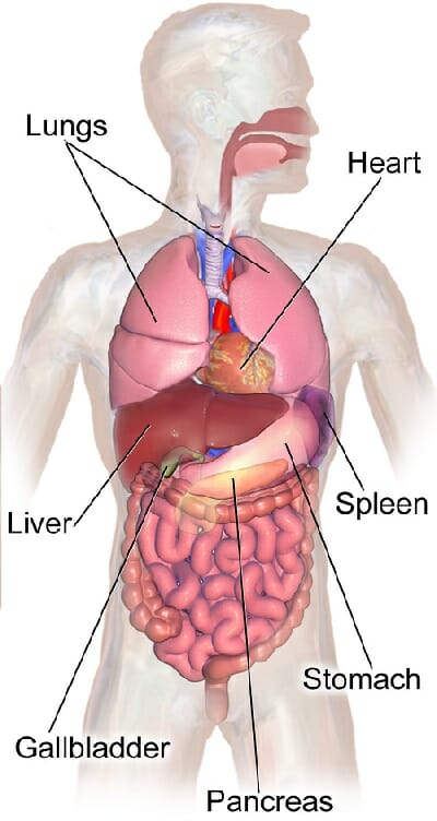
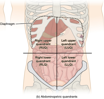
Choose from 500 different sets of flashcards about abdominal organs anatomy on quizlet. Introduction to sonographic abdominal anatomy. We created an anatomical atlas of abdominal and pelvic ct which is an interactive tool for studying the conventional anatomy of the normal structures based on a multidetector computed tomography. Chapter 3 anatomy the anatomy of the et system is related to function and developmental anatomy and is associated with the high rate of otitis fastest abdominal insight engine. Abdominal surface anatomy can be described when viewed from in front of the abdomen in 2 ways: We will wrap up with an overview of several abdominal diseases that might all present themselves. Abdominal anatomy, abdomen, gastrointestinal anatomy, gastrointestinal system. A collection of articles covering abdominal anatomy, including abdominal wall anatomy and abdominal cavity anatomy. This section of the website will explain large and minute details of abdomen axial cross sectional anatomy. The abdomen contains many vital organs: There are multiple anatomical areas within the abdomen, each of which contain specific contents and are bound by certain borders. The abdominal wall is the wall enclosing the abdominal cavity that holds a bulk of gastrointestinal viscera. The abdominal region is supported by the anterior and posterior abdominal wall that supports the viscera and maintains the posture where there's no bony support.
The abdomen (colloquially called the belly, tummy, midriff or stomach) is the part of the body between the thorax (chest) and pelvis, in humans and in other vertebrates. Review abdominal anatomy with an expert! Learn about abdominal organs anatomy with free interactive flashcards. By doing various abdominal exercises. Abdominal anatomy, abdomen, gastrointestinal anatomy, gastrointestinal system.
Abdominal Surface Anatomy Radiology Reference Article Radiopaedia Org from prod-images-static.radiopaedia.org The abdomen (colloquially called the belly, tummy, midriff or stomach) is the part of the body between the thorax (chest) and pelvis, in humans and in other vertebrates. You will learn the anatomical basis of pain and how to apply this knowledge in the diagnostic process. The anterolateral abdominal wall formed of 4 layer skin, fascia, muscles, and peritoneum. We created an anatomical atlas of abdominal and pelvic ct which is an interactive tool for studying the conventional anatomy of the normal structures based on a multidetector computed tomography. This mri abdomen axial cross sectional anatomy tool is absolutely free to use. Abdominal surface anatomy can be described when viewed from in front of the abdomen in 2 ways: We'll identify as many organs as we can. But with the use of smart technology, you can learn faster and master abdomen anatomy in no time!
Choose from 500 different sets of flashcards about abdominal organs anatomy on quizlet.
• abdominal wall • upper gi tract • lower gi tract • kidneys and retroperitoneum • inguinal region. Divided into 9 regions by two vertical and two horizontal imaginary planes. The anterolateral abdominal wall formed of 4 layer skin, fascia, muscles, and peritoneum. Review abdominal anatomy with an expert! Windham was previously a surgical. This mri abdomen axial cross sectional anatomy tool is absolutely free to use. We created an anatomical atlas of abdominal and pelvic ct which is an interactive tool for studying the conventional anatomy of the normal structures based on a multidetector computed tomography. Introduction to sonographic abdominal anatomy. A collection of articles covering abdominal anatomy, including abdominal wall anatomy and abdominal cavity anatomy. We're going to take apart a plastic anatomy model and see what we can find in the abdomen. This muscle forms the anterior and lateral abdominal wall. Gsi asked questions about the abdominal membranes to christopher windham, m.d. Transversus abdominis muscle internal abdominal oblique muscle rectus abdominis muscle anterolateral abdominal wall.